


Growth Plate Fractures
Children are unique in that their bodies are constantly changing and growing. The growth of bones is a result of active growth plates, and are only present in children. These growth plates are made of cartilage and, thus, more susceptible to injury compared to the surrounding bone. Pediatric orthopaedic surgeons are specialized specifically to address injuries to this vital area of a child’s anatomy.
The majority of injuries to the growth plate are relatively minor and will recover uneventfully. Even fractures that are displaced from their anatomic position often have the ability to self-correct through a process known as remodeling. Fractures that are displaced may require manipulation to reestablish the alignment of the bone within acceptable parameters. By maintaining the alignment of the bone within these guidelines, children often recover uneventfully.
Your surgeon may monitor your child’s recovery at a growth plate following a fracture that required manipulation to ensure that it has recovered uneventfully. Growth plate arrests are uncommon but may require additional treatments if they are diagnosed.
Buckle Fractures
Minor fractures are very common in growing children. Their bones are more flexible than adult bones and children are generally very active. Occasionally, as the result of minor trauma, a child may sustain a partial fracture of the bone, which is known as a buckle fracture. Buckle fractures can occur in any long bone, but they typically occur in the wrist or the leg.
These fractures derived their name from their appearance on x-ray as a “buckling” of the hard portion of the bone known as the cortex. The bone may or may not have a fracture that is completely through the bone. The fracture is usually stable in its position and does not typically require a physician to manipulate the bone.
These fractures often heal with simple protection and immobilization in a cast or a splint. Multiple studies have shown equivalent outcomes between casts and removable braces to treat buckle fractures of the wrist. These fractures generally require 3-4 weeks of protection for comfort and to minimize the risk of reinjury by another trauma.
Overall, there is a very low complication rate and children are expected to return to full recovery and activity with no long-term problems.
Upper Extremity Fractures
Wrist Fractures
Fractures of the wrist are common in children. They often occur when a child attempts to break her fall on outstretched hand. The two main bones of the wrist are the radius and ulna. Injuries range from minor “buckle fractures” to displaced fractures requiring the bone to be manipulated and possible hardware placement surgically. The fracture may or may not involve the child’s growth plate and have a grossly cosmetic deformity. There are strict parameters and acceptable guidelines for wrist fractures that your surgeon will discuss with you to determine whether further interventions will be necessary. Most fractures can be treated without manipulation and casting. If there is a block to overlying the fracture appropriately, surgery can remove the alignment block and pins are occasionally used to maintain the position of the bones until they heal. Most wrist fractures are healed uneventfully, and children recover well.
Forearm fractures
The forearm consists of two long bones called the radius and the ulna. These bones provide the framework and support for the forearm and the attachment points for muscles of the arm and hand.
Fractures of the forearm usually occur following a trauma and are confirmed with x-ray imaging. One or both of the bones may be broken. They may simply have a crack (known as a nondisplaced fracture), have a bend (known as a green stick fracture), or have a complete fracture with movement of the bones out of their natural position (displaced fracture). Severe injuries may also involve a cut in the skin that was made by the bone, exposing the bone to the external environment. This is known as an “open” or compound fracture.
Nondisplaced and mildly displaced fractures can often be treated with a long arm (above the elbow) fiberglass cast. Bones that are not aligned appropriately may require that your surgeon perform a maneuver (known as a reduction) to realign (set) the bones. This is typically done with sedative medicines. If your child’s injury is severe enough that it is unstable and will not maintain an acceptable alignment, then surgery is utilized to align the bones and often introduce temporary implants that will maintain the fracture alignment until there is an opportunity for the bones to heal. Once the bones have healed adequately, the implants are no longer necessary and your surgeon may give you the option to remove them at a later time, if desired.
Open fractures are often scrutinized closely, as there is an increased risk of bone infection due to the nature of the injury. These fractures are often explored and cleansed in the operating room to minimize the risk of a bone infection.
Once the fracture has healed, many patients notice that their arm is skinnier and perhaps has more hair than the other. This is due to a loss of muscle mass called “atrophy” and is a result of not using the muscles of the hand for several weeks. The size does return as the hand and arm are removed from immobilization and perform their normal functions. Increased hair stimulation is due to the rerouting of blood to nourish the injured area. As a side effect, hair follicles also receive extra nutrient stimulation to grow, causing the apparent increase in hair production.
Following removal of your cast it is important to begin moving your arm and elbow, following your physician’s recommendations. These motions include bending and flexing the elbow and wrist but also rotating the forearm to a palm up (supination) and palm down (pronation) position. If your child has difficulty with these motions, your surgeon may recommend the intervention of a therapist to aid in regaining mobility.
After the fracture has healed it continues to strengthen and reorganize itself to resemble the original bone. This process of “remodeling” typically takes 6 months and, although uncommon, slightly increases the risk of refracture at the original injury site. You should consult with your doctor about the risks of reinjury.
Elbow Fractures
Elbow fractures are a frequent cause of injury for the developing child. The bones of the elbow include the humerus, radius, and the ulna. The x-ray appearance of the elbow is difficult to interpret because it is constantly transitioning from cartilage to bone until early adolescence. Fractures remain in range from minor fractures that simply require immobilization to significant and complex fractures requiring surgical intervention and placement of implants. In general, minor elbow injuries heal without long-term consequence. Significant injuries may require additional rehabilitation and range of motion exercises to restore function. If the injury involves the growth plate, the fracture may need to be followed for several months to ensure that the bone has recovered adequately.
Supracondylar fractures are the most common site of the elbow fracture in children. Treatment depends on the level of displacement of the fracture. Treatment can range from casting without manipulation to surgical intervention requiring manipulation and pinning. Most fractures heal well, however, some may be complicated with injuries to nerves and blood vessels or limitation of motion.
Lateral condyle fractures are fractures of the joint surface. If the fracture is minimal this may be treated with simple casting, however most fractures of this kind will require surgery to realign the joint surface perfectly and secure in place with pins. Most of them are authorized for 6 weeks and then range of motion is begins. The patient also require therapy to address stiffness of the elbow following this injury.
Medial epicondyle fractures are injuries to the inside of the elbow. The major muscles of the forearm attached on the inside of the elbow. This type of injury is commonly seen in throwing athletes such as pitchers. This injury can be treated operatively or nonoperatively depending on the level of displacement and the athletic requirements of the patient.
Shoulder Fractures
Injuries of the shoulder may occur for a number of reasons. They most often involve the humerus bone near the shoulder. Many of these fractures can be treated with immobilization and observation. The shoulder is a very mobile joint and can accommodate some misalignment due to fracture. This misalignment often improves as a child ages through a process called remodeling. Functionally, the child generally resumes full range of motion and strength after the fracture has healed.
Clavicle Fractures
Clavicle fractures are common fractures that are most often seen in two populations of patients.
The youngest patients that are susceptible to clavicle fractures are newborn infants who fracture the clavicle as a result of the process of childbirth. These injuries occur in approximately 5 of every 1000 births. These injuries may be diagnosed on physical exam or x-ray. In general, children of this age have very flexible bones with exceptional healing potential. Approximately 1 in 10 children may have an injury to the nerves that control the arm. Most of the injuries will simply require close monitoring by your physician.
Older patients such as toddlers and adolescents typically sustain a clavicle fracture after trauma. It may be visible as a “lump” on the chest and is painful. Most fractures heal uneventfully without the need for manipulation or surgery. There are a few indications for surgery: 1) If the bone fragment penetrates the skin, 2) if there is significant shortening of the bone due to fracture, or 3) if the fracture is an unacceptable position. Relative, or conditional, indications for surgery include throwing athletes such as pitchers and quarterbacks, unacceptable cosmetic deformity, and shoulder weakness.
Most clavicle fractures are treated nonoperatively with a sling and lead to good results. The shoulder is typically immobilized for 2-3 weeks. On the 3rd week, the patient typically feels comfortable enough to discontinue use of the sling/shoulder immobilizer. By week 4, pain has generally resolved completely, and the patient has an improved to near normal range of motion. After week 4, the fracture continues to consolidate and strengthen and the patient typically has no symptoms, though there may be an underlying shoulder weakness from lack of use. Athletes typically return to play in 4-6 weeks, although it is recommended to refrain from contact sports for at least 8 weeks to minimize the risk of refracture.
Lower Extremity Fractures
Femur Fractures
The femur is the largest bone in the human body. Fractures can occur for a number of reasons in children of all ages. In infant children, it is common to involve a state caseworker as a matter of protocol to rule out nonaccidental injury. These injuries generally heal quickly and well and may require immobilization with a cast or soft harness. Injuries that involve the growth plate may inhibit normal growth of the leg, leading to a leg length discrepancy.
In older children, the fracture may be treated surgically or nonsurgically. Surgical indications focus on maintaining length and appropriate rotation of the leg. Implants that are placed are usually temporary and often have the option to be removed at a later time. The implants are biocompatible and designed to remain with the patient for their entire life span, but many surgeons offer the choice for elective removal once a fracture has completely healed.
Despite common belief, it is has not been clinically proven that these implants respond to changes in weather. The implants also should be magnetically neutral and not trigger metal detectors.
Leg (Tibia/Fibula)Fractures
The tibia and fibula compose the major bones of the lower leg. Fractures can occur for a number of reasons in children of all ages. In infant children it is common to involve a state caseworker as a matter of protocol to rule out nonaccidental injury. These injuries generally will heal quickly and well and may require immobilization with a cast. Injuries that involve the growth plate may inhibit normal growth of the leg leading to a leg length discrepancy.
In older children the fracture may be treated surgically or nonsurgically. Surgical indications focus on maintaining length and appropriate rotation of the leg. Implants that are placed are usually temporary and often have the option to be removed at a later time. The implants are biocompatible and designed to remain with the patient for their entire life span, but many surgeons offer the choice for elective removal once a fracture has completely healed.
Despite, belief it has not been clinically proven that these implants respond to changes in weather. The implants also should be magnetically neutral and not trigger metal detectors.
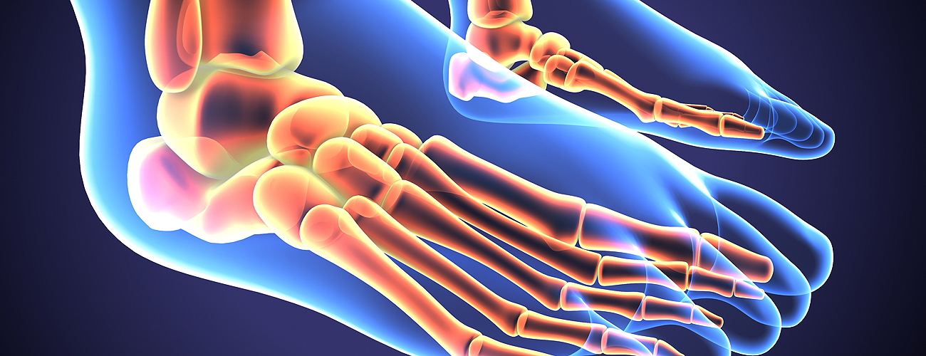
Ankle Fractures
Ankle fractures are common in young children. The major bones of the ankle include the tibia, fibula, and talus. Areas of the ankle may be susceptible to fractures due to weakened areas at the growth plate. The growth plate is made of cartilage and is more susceptible to injury than the surrounding bone. Fractures can occur through the growth plate of the fibula on the outside of the ankle or the tibia along the inside and front of the ankle. If the fracture has not moved it is often treated with immobilization. In the event that it has moved, manipulation and realignment by the physician may be required. Following the injury and recovery, the growth plate may require monitoring to ensure that it recovers adequately.